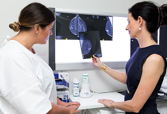
Breast tomosynthesis (3D mammography) is the latest and most advanced scientifically-proven technology for the early detection of breast cancer. Mammography uses extremely low levels of x-ray to create a digital image of the breast to be interpreted by our highly experienced radiologists, Dr Shane Connolly &/or Dr Caitlin Kapoor, who will report your study on the day. Any follow-up examinations required can then be arranged through our team.
It is very similar to a conventional mammogram only it creates very thin images of your breast from multiple angles and then renders this into a 3D image. This provides a superior depiction of breast tissue enabling more accurate differentiation between normal and abnormal breast tissue and allowing us to determine better what further investigation may be needed.
Your doctor may have also referred you for an ultrasound to be performed in conjunction with mammography. Breast ultrasound is used to provide additional information regarding the breasts and may clarify any uncertain areas seen on the mammogram. In women under the age of 35 years, ultrasound (rather than mammography) is usually performed as the initial investigation due to the increased density in breast tissue in those under 35.
The benefits of mammography regarding radiation exposure are deemed to far outweigh the risks and limitations, which include exposure to low levels of radiation. Mammograms can be difficult to interpret in younger women or those with dense breast tissue, and mammography can’t always detect every form of breast cancer.