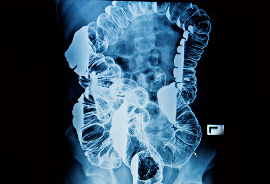
Fluoroscopy is an imaging technique that uses x-rays to obtain real-time moving images of the interior of an object. It allows us to assess the structure and function of a patient, so that the motion of swallowing, for example, can be watched. This is useful for both diagnosis and therapy. Fluoroscopy provides live moving pictures that can be observed by the radiologist during real time.
Barium studies and hysterosalpingography (HSG) form the main fluoroscopy studies at MD Imaging. The risks of fluoroscopy from minimal radiation exposure have already been considered by your referring practitioner, and at MD Imaging we adhere to the ALARA principle (as low as reasonably achievable) by using minimal screening times by highly experienced radiologists, Dr Shane Connolly and Dr Caitlin Kapoor, in order to reduce radiation exposure.
Barium studies include swallow, meal, follow through and enema. Risks of aspiration of Barium are exceedingly rare. Risks of enema include mucosal perforation but are also exceedingly rare.
HSG involves examination of the uterus and Fallopian tubes following speculum insertion through the vagina and radio-opaque contrast material through the uterus. Minor bleeding can occur following this procedure. Infection is rare.